Addressing massive talar bone loss and avascular necrosis through high-fidelity anatomical reconstruction.
Client:
Clinical Research Group
Date:
Feb 2025
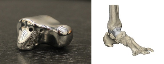
Total talar collapse, whether resulting from trauma-induced avascular necrosis (AVN) or severe osteoarthritis, presents one of the most significant challenges in reconstructive foot and ankle surgery. Traditional treatment pathways often defaulted to tibiotalocalcaneal (TTC) arthrodesis. However, fusion frequently results in a substantial loss of hindfoot mobility, altered gait mechanics, and secondary degeneration of adjacent joints.
The Total Talus Replacement application utilizes additive manufacturing to provide a motion-preserving alternative that restores joint height and anatomical alignment with extreme precision.
The process begins with high-resolution, thin-slice CT imaging of both the affected and contralateral (healthy) limb. By mirroring the healthy talus, our engineering team creates a digital "twin" that accounts for the patient’s unique articular geometry.
This application leverages Laser Powder Bed Fusion (LPBF) to print the device in medical-grade Titanium alloy (Ti-6Al-4V). Titanium is the material of choice for this application due to:
The 3D-printed talus is designed for a "press-fit" or "cemented" fixation depending on the quality of the surrounding bone stock. Because the implant is a geometric replica of the patient's original bone, the surgical team can minimize bone resection, preserving as much native vascularized tissue as possible.
Observed Clinical Benefits Include:
Clinical validation through multi-centered studies suggests that patient-specific talar replacements provide superior functional scores (AOFAS) compared to traditional fusion. By combining regulatory-aware design with evidence-based innovation, this application represents the gold standard in limb-salvage technology.
.png)
Join a trusted ecosystem dedicated to the advancement of 3D-printed implant technologies. We transform complex anatomical challenges into clinical successes through evidence-based innovation and regulatory transparency.

