The clinical success of modern orthopedic hardware is fundamentally defined by the Biological-Mechanical Interface. While traditional "solid" implants rely on surface coatings for secondary stability, next-generation additive manufacturing allows for a monolithic porous architecture. The primary variable in this equation is Lattice Density, which dictates the balance between structural load-bearing and osteoblastic infiltration.
The Architecture of Osseointegration
Osseointegration is not merely a surface event; it is a three-dimensional biological migration. To facilitate this, the lattice must be engineered with specific geometric constraints that mimic the host environment.
- Interconnected Porosity: Unlike traditional porous coatings, 3D-printed lattices are 100% interconnected. This allows for the unimpeded flow of nutrients and the deep penetration of vascular networks, which are the precursors to mature bone formation.
- Mean Pore Diameter: Research has identified an "Optimal Window" between 600 and 800 microns. Pores below this threshold restrict cell signaling and lead to fibrous tissue rather than bone, while pores exceeding this limit compromise the fatigue life of the titanium.
- Surface Micro-Topography: The "as-printed" surface of Laser Powder Bed Fusion (LPBF) titanium naturally possesses a sub-micron roughness. This micro-texture is critical for initial protein adsorption and the immediate "scratch fit" stability required in the operating room.
Mechanical Biomimicry and Stress Distribution
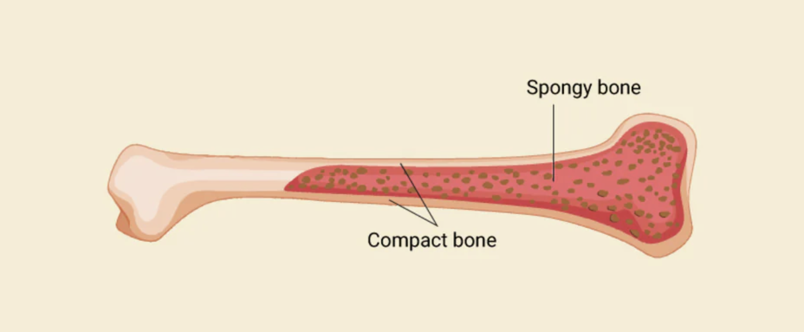
One of the most significant advantages of adjustable lattice density is the ability to match the Elastic Modulus of human bone. Traditional solid titanium is significantly stiffer than cortical or cancellous bone, leading to a phenomenon known as Stress Shielding.
- Load Sharing: By reducing the density of the implant's core, we lower its global stiffness. This ensures that the physiological load is shared with the host bone, stimulating natural remodeling and preventing bone resorption.
- Structural Integrity: Finite Element Analysis (FEA) is utilized to ensure that even at lower densities, the lattice can withstand millions of loading cycles without structural deformation.
- Vascularization Pathways: The density of the lattice is strategically varied to create "highways" for blood vessel growth, ensuring the long-term viability of the bone within the scaffold.
Clinical Outcomes and Data-Driven Design
The transition to optimized lattice structures has resulted in measurable improvements in secondary stability and implant longevity. By correlating specific density parameters with patient-reported outcome measures (PROMs), we have moved beyond "one-size-fits-all" manufacturing.
- Secondary Stability: Longitudinal imaging confirms that bone in-growth into these porous structures provides a mechanical bond that is significantly more resilient than traditional cement or grit-blasted surfaces.
- Reduced Revision Rates: By matching the biomechanical properties of the native anatomy, we reduce the primary drivers of implant loosening and mid-term failure.
.png)