The realization of a patient-specific implant relies on a seamless "Digital Chain of Custody" that bridges the gap between clinical radiology and advanced additive manufacturing. This workflow is a highly specialized engineering sequence designed to ensure that the final 3D-printed titanium device matches the patient's unique anatomy with sub-millimeter precision.
Phase I: High-Fidelity Anatomical Data Acquisition
The foundation of every custom implant is high-resolution medical imaging. Unlike off-the-shelf components, the patient-specific process begins with a specialized scanning protocol tailored for metal-bone differentiation.
- Thin-Slice CT Imaging: We utilize non-interpolated, thin-slice CT data (typically 0.625mm or less) to capture the intricate geometry of bone defects or articular surfaces.
- Metal Artifact Reduction (MAR): In revision cases where existing hardware is present, advanced MAR algorithms are applied to prevent digital "noise" from obscuring the bone-implant interface.
- Anatomical Segmentation: Using AI-assisted software, engineers isolate the target anatomy from surrounding soft tissue, creating a high-fidelity 3D volume that serves as the "digital twin" of the patient.
Phase II: Virtual Surgical Planning and FEA
Once the anatomical model is finalized, the engineering team collaborates with the surgeon to define the implant's boundaries and functional requirements.
- Boolean Subtraction: The implant is designed to perfectly complement the patient’s defect, ensuring a "lock-and-key" fit that maximizes surface contact area.
- Finite Element Analysis (FEA): Before a single grain of titanium is fused, the design undergoes rigorous computational stress testing. We simulate physiological loads—such as walking or stair climbing—to identify and eliminate potential stress concentrations.
- Screw Path Optimization: For complex reconstructions, screw trajectories are pre-planned digitally to ensure maximum purchase in the highest-density bone while avoiding critical neurovascular structures.
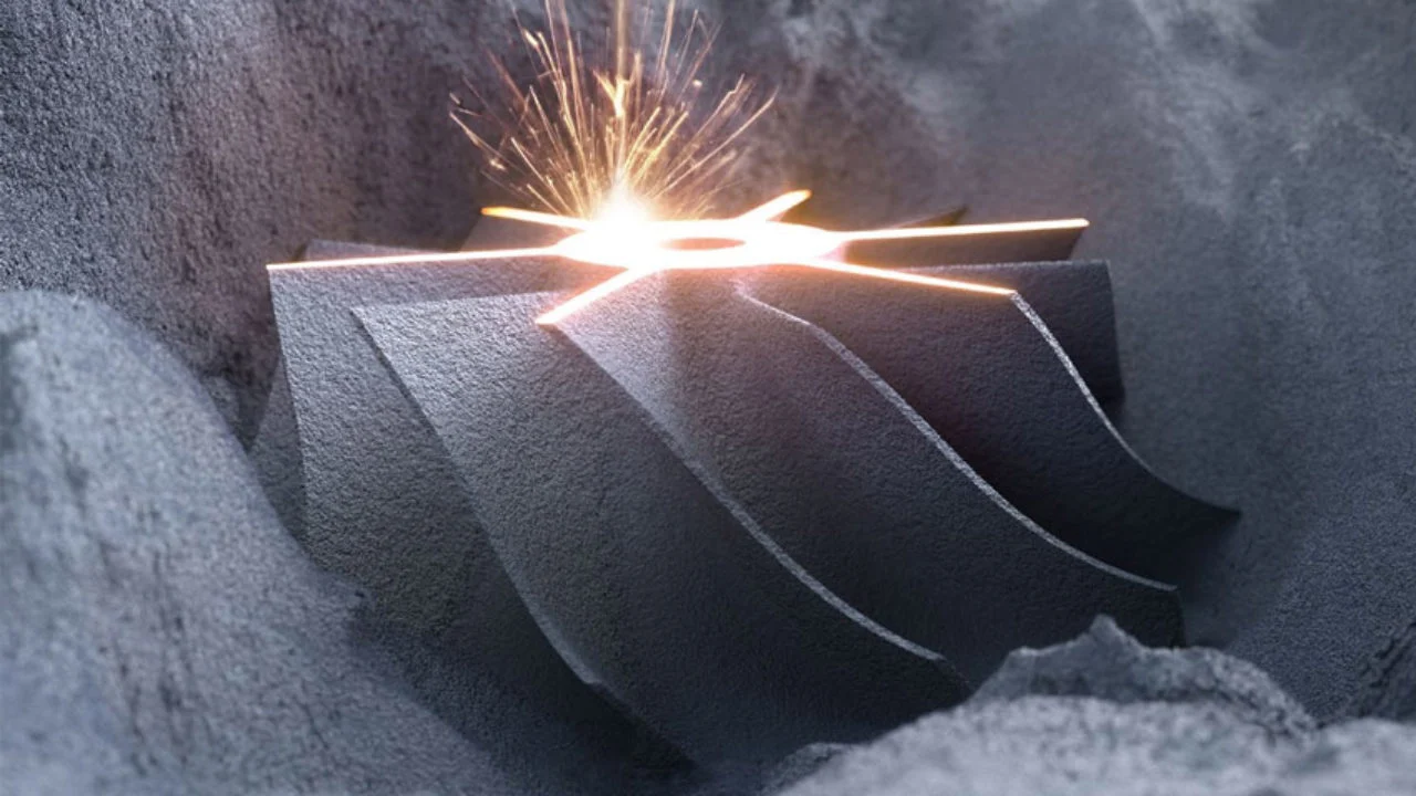
Phase III: Laser Powder Bed Fusion (LPBF)
The transition from digital to physical occurs within a controlled inert environment using medical-grade Titanium alloy (Ti-6Al-4V).
- Monolithic Printing: The device is grown layer-by-layer, allowing for the integration of complex internal lattices and solid structural members into a single, seamless component.
- Thermal Management: Specialized support structures are engineered to act as heat sinks during the print process, preventing geometric warping and ensuring the final device matches the digital design within microns.
- Digital Verification: Post-print, the device is laser-scanned and compared back to the original STL file to verify dimensional accuracy before moving to final processing.
Phase IV: Post-Processing and Clinical Delivery
The final stage involves transforming the raw printed part into a sterile, surgical-grade medical device.
- Surface Refinement: Articular surfaces undergo high-precision polishing to achieve a mirror finish, while non-articular surfaces retain a specific micro-roughness to encourage biological fixation.
- De-Powdering and Cleaning: Proprietary ultrasonic cleaning protocols ensure that all residual metal particles are removed from complex, porous internal geometries.
- Sterilization and Tracking: Each implant is uniquely serialized, providing a transparent record from the initial CT scan through to the moment of implantation in the operating room.
.png)